
The axial skeleton is the central framework of the human skeleton, consisting of bones located along the body’s central panel. It includes the following components:
- Skull:
8 cranial bones and 14 facial bones (forming the face). - Vertebral Column:
Made up of cervical, thoracic, lumbar, sacral, and coccygeal vertebrae, i - Ribcage:
Consisting of true ribs, false ribs, and floating ribs, it safeguards the vital organs in the chest and facilitates breathing. - Hyoid Bone:
Located in the neck, it’s crucial for tongue and neck muscle attachment, aiding in speech and swallowing.
Significance:
- Support and Protection:
The axial skeleton serves as the central structural support for the body, allowing us to maintain an upright posture. It also protects vital organs:
– The skull guards the brain, a crucial organ.
– The ribcage protects vital organs like the heart and lungs from external trauma/ injury.
– The vertebral column safeguards the spinal cord, the central nervous system.
– The hyoid bone provides support for the neck and tongue muscles. - Facilitation of body Movement:
The axial skeleton is primarily concerned with support and protection, it also plays a role in movement. The vertebral column, in particular, allows for various degrees of flexibility and motion like bending, twisting, and rotation. The skull’s bones involve in articulation for speech, while facial bones are essential for facial expressions. - Hematopoiesis:
Red bone marrow present within these axial skeleton bones like sternum produces red blood cells, white blood cells, and platelets, which are essential for overall health. - . Crucial in Development: During embryonic development, it provides a foundation for the appendicular skeleton (limbs and girdles) and other bodily structures.
Components of the Axial Skeleton
A. The Skull
- Cranial bones [8]
- Facial bones [14]
B. The Vertebral Column
- Cervical vertebrae [7]
- Thoracic vertebrae [12]
- Lumbar vertebrae [5]
- Sacrum [5]
- Coccyx [4]
C. The Ribcage has 12 pairs
- True ribs [7 pairs ]
- False ribs [3 pairs ]
- Floating ribs [2 pairs ]
D. The Hyoid Bone
Functions of the Axial Skeleton
- Protection of vital organs.
- Plays an important role in maintaining body posture.
- The vertebral column helps in Movement.
- skull plays an important role in articulation and facial expressions
- axial skeleton also significant in blood cell production
How does axial skeleton develop?
During the early phase of embryonic development, the human embryo consists of three primary germinal layers: Ectoderm, Mesoderm, and Endoderm.
- Formation of Mesoderm begins then the mesoderm further differentiates into somite and somite Formation occurs [ somite is a longitudinal series of block like segments into which the mesoderm, the middle layer of tissue, on either side of the embryonic spine, becomes divided. Collectively, the somite constitute the vertebral plate.]
- These somite give rise to various musculoskeletal structures, including the axial skeleton.
- Development of the Skull: The skull is the most complex part of the axial skeleton. It develops through a combination of membranous ossification and endochondral ossification processes:
- Membranous ossification: Some of the skull bones, such as the flat bones of the skull (parietal and frontal bones) are formed directly from condensed mesenchymal tissue through a process called intramembranous ossification.
- Endochondral ossification: Some other parts of the skull, like the base of the skull and facial bones, develop from cartilage models through endochondral ossification. Cartilage is then gradually replaced by bone tissue.
Formation of the Vertebral Column:
The vertebral column develops from somites located along the length of the embryo. These somites differentiate into sclerotomes, which give rise to the vertebrae. The vertebrae begin as cartilaginous structures and gradually ossify over time.[edited ]
Ribcage Development:
The ribs and sternum (breastbone) form from the lateral plate of the mesoderm. The ribs initially develop as cartilaginous structures which undergo ossification to become bony ribs. The sternum also forms a cartilaginous structure and later gradually ossified.[edited]
Development of the Hyoid Bone:
The hyoid bone itself is unique in that it does not articulate with any other bones. It forms from the paired lateral cartilage elements known as Reichert’s cartilage, later fuse in the midline during embryonic development.
How the axial skeleton evolves over the different stages of life:
During Infancy and Childhood:
- Growth and Ossification: During infancy and childhood, the axial skeleton continues to grow and develop like the bones of the skull, vertebral column, and ribcage undergo further ossification.
- Fontanel Closure of head: Fontanels, the soft spots on an infant’s skull, gradually begin to close as the cranial bones start to fuse together. This process leads to the development of the adult skull.
- Spinal Development: The vertebral column elongates, and the individual vertebrae continue to ossify. As a result, a child’s spine becomes more developed and better able to support the body’s increasing weight.
During Adolescence:
Adolescence is generally an age of rapid growth. The axial skeleton adapts to accommodate the increased height and weight of the individual. This phase is significant for the development of the vertebral column, which may experience changes in the curvature of the bone.
Skeletal Maturation: Many bones in the skull, vertebral column, and ribcage continue to mature and solidify with an increase in bone density and the bones become denser and stronger.
During Adulthood
The axial skeleton is more stable in early adulthood, with ongoing bone remodeling processes. New bone tissue[ osteoblast ] replaces old bone tissue to maintain strength.
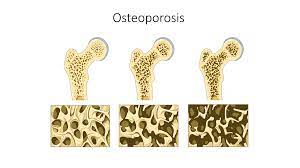
Degenerative Changes: As an individual enters into old age, degenerative changes may also begin to occur. For example, The intervertebral discs in the spine can lose water content and resilience, leading to decreased height and increased risk of conditions like herniated discs. Osteoporosis is a condition characterized by a decrease in bone density. This condition can affect the axial skeleton, leading to an increased chance of fractures.
Also In old age, the axial skeleton continues to undergo degenerative changes with ages. These changes lead to further loss of bone density, and the development of osteoarthritis in the spine and ribcage joints.
- There is increased Fracture Risk with reduced bone density and strength, especially hip and vertebral fractures, which becomes more pronounced in the elderly.
Throughout an individual’s life, proper nutrition, exercise, Diet and healthcare play an important role in maintaining the health and strength of the axial skeleton. Regular physical activity, a balanced diet rich in calcium and vitamin D, and lifestyle choices can help avoid some of the age-related changes and reduce the risk of skeletal disorders.
Common Disorders and Conditions
A. Osteoporosis
B. Scoliosis
C. Skull fractures
A. Osteoporosis: Osteoporosis is a skeletal disorder characterized by a decrease in bone density and structural deterioration of bone tissue[ osteoblast ] resulting in weak bones that are more exposed to risk of fractures, even with minor trauma.
B. Scoliosis:
Scoliosis is a medical condition that is characterized by an abnormal lateral (sideways) curvature of the spine. The spine may form an “S” or “C” shape when viewed from the front or back and may have various causes, including congenital factors, neuromuscular conditions, etc.
C. Skull Fracture:
Skull fractures are the breakage of one or more bones in the skull. These fractures can vary in severity, from a hairline crack to more severe fractures of multiple bone pieces. resulting from falls, sports accidents, Road traffic accidents, or assaults.
Diagnostic Techniques and Medical Imaging
- X-ray
- CT-scan
- MRI scan
X-rays (Radiography):
- X-rays are a form of electromagnetic radiation. When directed at the body, they are able to pass soft tissues but are absorbed by denser structures like bones, leaving an image on a detector
- X-rays are commonly used to visualize bone fractures, including those of the skull, ribs, vertebral column, and other axial bones.
- They can assess in diagnosing arthritis and joint conditions by showing joint space narrowing and bony degenerative changes.
- In dentistry, X-rays are useful to examine the teeth and jaw, which are part of the axial skeleton.
CT Scans (Computed Tomography):
- CT scans work by using a series of X-ray images taken from different sides and angles to create detailed cross-sectional images of the body offering completely superior details in both bones and soft tissues compared to X-rays.
- It provides detailed images of bone structures, making them helpful in assessing complex fractures, bone tumors, and bone abnormalities.
- Valuable in evaluating spinal disorders, such as herniated discs, spinal stenosis, and vertebral anomalies.
- Trauma injury Evaluations are often done by CT scan in trauma cases to quickly assess head, face, and spine injuries.
MRI Scans (Magnetic Resonance Imaging):
- MRI uses strong magnetic fields and radio waves to generate detailed images of the body’s internal structures and not by ionizing radiation.
- It is applicable in soft tissue assessment making it valuable for diagnosing conditions affecting the spinal cord, nerves, muscles, and ligaments in the axial skeleton.
- MRI is the primary imaging modality for evaluating brain and spinal cord conditions, including tumors, multiple sclerosis, and spinal cord injuries.
- Also, MRI angiography can assess blood vessels in the brain and neck, which are part of the axial skeleton.
Treatment and Management
- Orthopedic interventions
- Surgical procedures
- Physical therapy
Treatment and management strategies for conditions related to the axial skeleton, such as those mentioned earlier (e.g., fractures, scoliosis, herniated discs, osteoporosis), often involve a combination of approaches, including orthopedic interventions, surgical procedures, and physical therapy. Here’s an overview of how these treatment options are used:
A. Orthopedic Interventions
Orthopedic interventions refer to non-surgical medical procedures and treatments provided by orthopedic specialists, who are experts in musculoskeletal conditions.
Applications:
- Conservative Fracture Management:
For non-severe fractures of axial bones, immobilization with casts, braces, or splints is done. - Orthotic Devices:
Orthotic devices, such as custom-made shoe inserts or braces, to address conditions like scoliosis or provide support for weakened or injured areas are used. - Medical Management of Osteoporosis:
Orthopedic specialists often work with patients to manage osteoporosis through medications, lifestyle modifications, and fall prevention strategies.
B. Surgical Procedures:
Surgical procedures involve physically addressing and correcting anatomical issues related to the axial skeleton through surgical techniques.
Applications:
- Fracture Repair:
Severe fractures involving multiple bone fragments or dislocated vertebrae, may require surgical reduction like open internal fixation. - Scoliosis Correction:
In severe scoliosis cases, the spine’s curvature is significantly affecting health or function. Procedures like spinal fusion are used to straighten and stabilize the spine are used . - Herniated Disc Removal:
In cases of persistent pain or neurological symptoms due to herniated discs, surgical procedures like discectomy or microdiscectomy may be performed to remove or repair the affected disc.
C. Physical Therapy:
Physical therapy involves different techniques and exercises to improve mobility, strength, and function, and reduce pain.
Applications:
- Rehabilitation after Surgery:
In many post-operative recoveries of axial skeleton-related surgeries physical therapy is beneficial to regain strength and mobility. - Conservative Treatment:
For conditions like scoliosis or non-severe fractures, physical therapy may be prescribed to strengthen supporting muscles, improve posture, and manage pain. - Pain Management:
Application of heat, cold, electrical stimulation, and manual techniques to alleviate pain and improve mobility.
In summary, the axial skeleton, particularly the skull and bones, has cultural significance as symbols in various traditions and as artistic motifs. Furthermore, the historical study of skeletal anatomy has shaped our understanding of the human body and its place in the natural world, contributing to scientific and medical progress over centuries