
Introduction
The human body is a perfect creation of nature, finely designed with an array of proper systemic mechanisms that ensure its survival. Among its many structures, the ribcage stands as strong evidence of nature’s genuineness in protecting vital organs. Often described as the body’s protective cage, the ribcage is an essential shield for the heart and lungs, offering both structural support and protection against external traumas. In this article, we will explore the human ribcage’s anatomy, function, significance, Injury, and management.
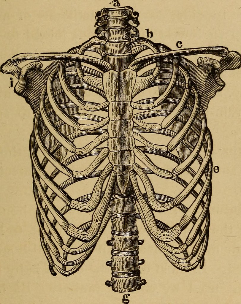
Human Ribcage
The human ribcage is a framework of bones that encloses the thoracic cavity, providing support and protection to the organs. It is composed of 12 pairs of ribs, collectively named the thoracic cage. It is situated between the spine and the sternum (breastbone). These ribs are divided into three categories Ribs. The ribs are the elongated bones that form the primary structure of the ribcage. Each rib consists of a shaft and two ends:
- True Ribs: The first seven pairs of ribs in the cage are true ribs which directly attach to the sternum with costal cartilage, forming a rigid and stable structure.
- False Ribs: Ribs 8, 9, and 10 are called false ribs which are indirectly connected to the sternum through cartilage that links them to the seventh rib.
- Floating Ribs: The remaining two pairs of ribs, 11 and 12, are floating ribs. They are not attached to the sternum at all and are free-floating in the lower part of the ribcage.
Anatomy of the Human Rib
- Head: The head of the rib is mainly the flattened portion that connects with the vertebral column. There are two facets on the head that connect to the corresponding thoracic vertebrae, creating the costovertebral joints.
- Neck: The neck is a narrow portion just beyond the head.
- Tubercle: The tubercle is a small, knob-like projection on the posterior surface of the rib that joins with the transverse process of the corresponding thoracic vertebra.
- Shaft (Body): The ribbed shaft is a long, curved portion extending from the neck to the anterior of the ribcage.
- Costal Cartilage: Each rib, except for the last two pairs (11,12), connects to the sternum via costal cartilage. These cartilages provide flexibility and allow for slight movement during breathing.
Sternum (Breastbone): The sternum is a flat, elongated bone located in the center of the anterior chest. It consists of three parts:
- Manubrium: The uppermost part of the sternum,
- Body (or Gladiolus): The elongated central portion of the sternum, which connects to the costal cartilages of the true ribs.
- Xiphoid Process: The small, pointed part at the inferior (lower) end of the sternum.
Vertebral Column (Spine):.The vertebral column, or spine, is the bony structure that runs along the posterior aspect of the ribcage. Ribs only connect to the 12 thoracic vertebrae but the spine can be divided into five regions. Cervical (neck), thoracic, lumbar (lower back), sacral (pelvic region), and coccygeal (tailbone).
Function of the Ribcage
The ribcage has several critical roles in maintaining the body’s overall well-being:
- Protection of vital organs: The primary function is to protect the vital organs within the thoracic cavity, mainly the heart and lungs. Without this protective cage, our vital organs would be vulnerable to injury, making life exceedingly precarious. The ribs act as a natural barrier against external forces or trauma, such as impacts or compressive loads, which could otherwise damage these essential organs.
- Support to upper body: The ribcage provides structural support to the upper body too. It forms a strong foundation for the attachment of muscles, allowing for various respiratory and upper-body movements, such as breathing and arm movements.
- Foundation of respiration: The ribcage is essential for the mechanics of breathing. During inhalation, the ribs elevate and expand up and outside, increasing the thoracic volume. This expansion creates a vacuum in the lungs, pulling in air. During exhalation, the ribs return to their resting position, helping in the expulsion of air.
- Blood Cell Production: Red bone marrow present within the cavities of the ribs, plays a vital role in the production of red and white blood cells, contributing to the body’s immune system and oxygen transport.
So, human ribcage is more than just a structural framework; it is a fundamental component of our survival. Without this protective cage, our vital organs would be vulnerable to injury, making life exceedingly precarious. Its role in respiration ensures that our bodies receive the oxygen required for energy production. It also helps to remove carbon dioxide, a waste product of metabolism.
Inhalation (Inspiration):
During inhalation, the ribs play an important role in expanding the thoracic cavity generating pressure inside, and facilitating the intake of air into the lungs:
Role of ribs and intercostal muscles in inhalation and exhalation.
- External Intercostal Muscles: Positioned between the ribs, the external intercostal muscles contract, causing the ribs to move upward and outward. This action enlarges the ribcage, expanding the thoracic cavity in multiple directions.
- Diaphragm Contraction: Also the diaphragm, a dome-shaped muscle situated at the base of the ribcage, contracts and flattens. This contraction increases the vertical dimension of the thoracic cavity.
- Thoracic Volume Expansion: As a result of the combined action of the external intercostal muscles and the diaphragm leads to an expansion of the thoracic cavity reducing the pressure within the thoracic cavity, and creating a vacuum. Consequently, air is drawn into the lungs through the respiratory tract, moving from areas of higher atmospheric pressure outside the body enabling the intake of oxygen and the exchange of gases.
Exhalation (Expiration):
During passive exhalation at rest, the ribcage and intercostal muscles primarily play a role in relaxation:
- External Intercostal Muscles: Once the air is inspired in full volume these muscle contracts and the internal lung pressure increases than outer atmospheric pressure after an exchange of gases with oxygen and carbon dioxide as a result these muscles relax, allowing the ribs to return to their resting position. This reduces the space and also within the thoracic cavity.
- Diaphragm Relaxation: The diaphragm relaxes, returning to its dome-shaped position. This relaxation decreases the vertical dimension of the thoracic cavity.
- Air Expulsion: As the ribcage and diaphragm relax, This elevated pressure and decreased space causes air to be expelled from the lungs through the respiratory tract, resulting in passive exhalation.
During heavy physical activity, additional muscles contribute to deep breathing like :
- Internal Intercostal Muscles
- Abdominal Muscles: Abdominal muscle contracts and raises abdominal pressure, pushing the diaphragm upward and assisting in the expulsion of air from the lungs.
These combined mechanisms enable efficient inspiration and expiration, supporting the body’s respiratory needs.
Development of Ribs from infancy to adulthood.
Breathing and ribs during Infancy
In infancy, the ribcage is soft and flexible due to the presence of cartilage in the developing ribs also relatively small and more cylindrical in shape and oriented horizontally, which is partly due to the infant’s lying-down position during this stage of life. The cartilage provides some degree of flexibility, which allows for the expansion of the ribcage during breathing. It assists during the passage of the infant through the birth canal during delivery. Moreover, diaphragm and abdominal muscles are in used for breathing
During Childhood and Adolescence
As children grow and develop, their ribcage undergoes different changes. They gradually ossify, a broader upper portion, and a narrower waistline, replacing the cartilage with bone. This makes the ribcage more rigid and stable over time. The ribcage expands both horizontally and vertically as the child grows. These ribs elongate and the spaces between them (intercostal spaces) widen, increasing the ribcage’s overall size. As the ribcage grows and matures, children gradually transition from predominantly diaphragmatic breathing to thoracic (chest) breathing. The ribcage becomes more involved in the expansion and contraction of the thoracic cavity during breathing.
During Adulthood
Fully Developed Structure: By adulthood, the ribcage has reached its full structural development. The ribs are fully ossified, and barrel-shaped with increased volume capacity. These provide maximum stability and protection to the thoracic organs which play a central role in inhalation and exhalation.
In summary, the ribcage undergoes significant transformations from infancy to adulthood. These changes involve the replacement of cartilage with bone, growth in size and proportion, alterations in shape, and shifts in breathing mechanics. The fully developed adult ribcage provides stability, protection, and optimal support for the respiratory system, facilitating efficient breathing and maintaining the health of the thoracic organs.
Discuss the role of the ribcage in speech and sound production.
Common Ribcage Injuries and Conditions in Thoracic cavity
- Rib Fractures: Rib fractures can result from trauma such as falls, Road traffic accidents, sports injuries, or direct blows to the chest leading to symptoms like Pain with breathing or movement, touch at the site of the fracture, chest wall tenderness, and potentially difficulty breathing.
- Treatment: Rest, pain management, and sometimes bracing or taping of the affected area. Severe fractures may require surgery.
2. Costochondritis: Inflammation of the cartilage that connects the ribs to the sternum, often due to injury, overuse, or infection leading to symptoms like Sharp chest pain, at the joints of the ribs and sternum. Exacerbation by deep breathing or movement may be seen.
- Treatment: Rest, anti-inflammatory medications, physical therapy, or local injections.
3. Rib Dislocation or Subluxation: Dislocation occurs when a rib slips out of its normal anatomical position, typically due to trauma. Subluxation is a partial dislocation with similar symptoms like Severe pain and discomfort, especially with breathing and movement.
- Treatment: An attempt to manually reposition the dislocated rib can be done by an expert medical person. Rest and pain management are necessary.
4. Flail Chest: A critical condition with multiple adjacent rib fractures in multiple places, leading to detachment of a segment of the chest wall.
- Treatment: Usually involves stabilizing the flail segment with bracing or surgery to repair the fractured ribs.
6. Pectus Carinatum (Pigeon Chest): Congenital chest wall deformity characterized by a protrusion of the sternum, creating a “pigeon chest” appearance.
- Treatment: Bracing or surgical correction may be considered in severe cases.
It’s important to note that any chest injury or persistent chest pain should be evaluated by a healthcare professional, as some conditions can be serious and may require medical intervention. Proper diagnosis and treatment are essential for a full recovery and to prevent potential complications.
Maintaining Ribcage Health
1. Maintaining proper posture helps support the natural alignment of the ribcage and spine. Sit and stand up straight which can put unnecessary stress on the ribcage.
2. Engage in Regular Exercise like cardiovascular exercises like jogging, swimming, cycling, and strength training into your fitness routine.
3. Practicing deep breathing exercises to enhance lung capacity and strengthen the respiratory muscles.
4. Excess body weight can result in straining the ribcage and chest muscles. So healthy weight through a balanced diet and regular exercise is necessary.
5. Avoid Smoking as it can weaken the ribcage by affecting lung health and increasing the risk of respiratory conditions.
8. To prevent rib fractures or injuries, take safety precautions during sports or activities that carry a risk of chest impact.
9. Consume a well-balanced diet rich in nutrients that support bone health, such as calcium and vitamin D.
10 . Regular Check-ups: — Regular check-ups with your healthcare provider to assess overall health.
Conclusion
Remember that maintaining a healthy ribcage is not only important for physical health but also contributes to overall well-being and the efficient functioning of the respiratory system. Consult with a healthcare professional or fitness expert for personalized advice and exercise recommendations based on your individual needs and health status. As we contemplate the remarkable role of the ribcage in protecting our most vital organs and enabling the breath of life, let us not only appreciate its silent, steadfast guardianship but also recognize the importance of actively maintaining its health.
From good posture to regular exercise and mindful breathing, the choices we make today can impact the strength and resilience of our ribcage for a lifetime. So, let us embrace a holistic approach to our well-being, cherishing the intricate design of our body’s protective cage and nurturing it with the care it deserves. In doing so, we honor the gift of life and the extraordinary mechanisms that sustain it, encouraging a healthier, more harmonious existence.
Best👏