
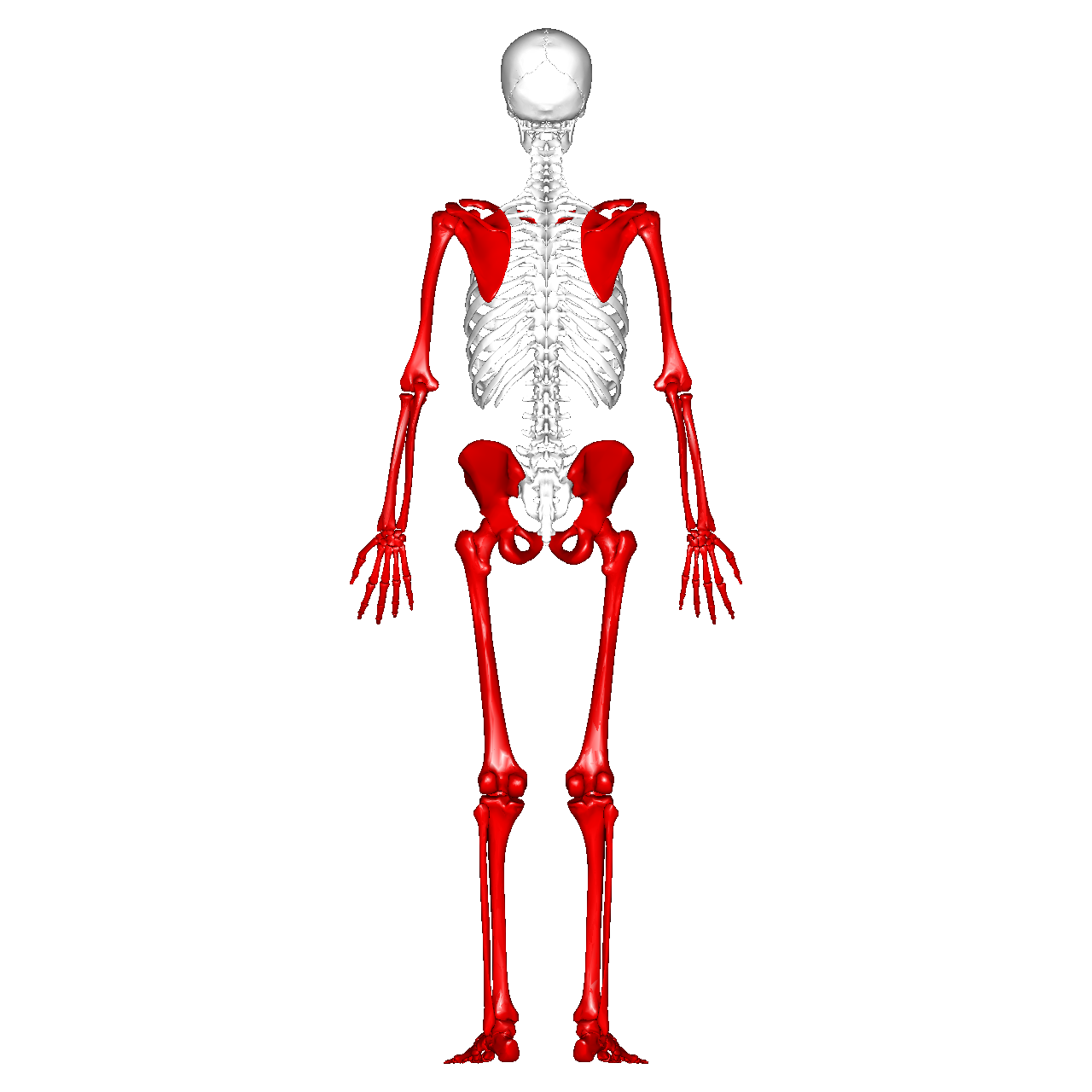
There are two major divisions of the human frame, which are the Appendicular skeleton and the axial skeleton. It includes all the bones that form the limbs and the girdles that connect the limbs to the axial skeleton. In brief, the appendicular framing is responsible for our mobility and the manipulation of objects. The appendicular structure mainly consists of the upper extremities and lower extremities which are the pelvis and shoulder girdle.
Components of Appendicular Skeleton
The appendicular skeleton is made up of the bones in the upper and lower limbs. This includes the shoulder girdle (clavicle and scapula), the arms (humerus, radius, ulna), the hands including carpals, metacarpals, and phalanges, the pelvic girdle (pubis), the legs including femur, tibia, fibula, and the feet including tarsals, metatarsals, and phalanges. These components are joined together by tendons and ligaments which make the human body a beautiful creation whose major role is facilitating the movement of the body and a wide range of activities like walking and performing fine motor tasks with our hands.
A total of 206 bones in the adult human body, 80 bones form the Axial skeleton, and a total of 126 bones form the appendicular skeleton. The bones that contribute to the appendicular skeleton include the bones of the hands, feet, upper extremity, lower extremity, shoulder girdle, and pelvic bones.
Here are mentioned names of all 126 bones of the appendicular skeleton.
Upper Limb
- Clavicle(1*2)
- Scapula(1*2)
- Humerus(1*2)
- Radius (1*2)
- Ulna(1*2)
- Metacarpal(5*2)
- Phalanges(14*2)
- Carpal Bones (8*2)
– Scaphoid
– Lunate
– Triquetrum
– Pisiform
– Trapezium
– Trapezoid
– Capitate
– Hamate
Lower Limb
- Pelvic girdle
– Ilium
– Ischium
-Pubis - Femur(1*2)
- Tibia(1*2)
- Fibula(1*2)
- Tarsals(5*2)
– Talas
– Calcaneus
– Cuboid
– Medial, intermediate, and lateral cuneiform
– Navicular - Metatarsals(5*2)
- Phalanges (14*2)
Development of the Appendicular Skeleton
The development of the appendicular skeleton is a journey that begins from the period of embryonic stage and continues throughout an individual’s life. Key points in this developmental process include:
- Embryonic Development
- Neonatal Growth
- Age Changes
Embryonic Development: During the period of embryonic development, a baby undergoes various changes and development of the body in structural anatomy. In the case of the Appendicular Skeleton the limb buds form, which will eventually become the upper and lower limbs. There are three layers of Embryonic tissue which are Ectoderm, Mesoderm, and Endoderm. The mesoderm, a middle layer of embryonic tissue, gives rise to bone-forming cells that later differentiate into the bones of the limbs.
Neonatal growth: After proper embryonic development, the appendicular skeleton undergoes substantial growth in a postnatal period known as neonatal growth with long bones lengthening through the process of endochondral ossification. Growth plates that are present at the ends of long bones allow increments of bone length as an individual matures. This growth continues into adolescence and is critical for achieving the full range of motion and strength in the limbs.
Age changes: As the body grows older and older, the appendicular skeleton of an individual experiences changes such as a decrease in bone density, which can lead to conditions like osteoporosis. Additionally, the joints may undergo wear and tear, leading to conditions like osteoarthritis.
Functions of the Appendicular Skeleton
The appendicular skeleton performs several critical functions that are integral to human’s daily activities. These functions can be broadly categorized as follows:
- Facilitating Movement: The appendicular skeleton is hugely responsible for movement, allowing us to walk, run, swim, eat, and perform various daily activities. The joints at the shoulder, elbow, hip, and knee allow us flexion, extension, rotation, and other complex movements that ease the living of life and make it easier.
- Stability and Support: Different bones like bones of pectoral and pelvic girdles provide a stable foundation for the attachment of the limbs to the axial skeleton. This stability is essential for maintaining balance and performing movements in daily life.
- Manual Functions: The upper limbs, especially the design of the hand and fingers, are fine for grasping, holding, and manipulating objects. The combined articulation of multiple hand bones, along with the flexibility of finger joints, offers a wide range of manual dexterity.
- Supports Body Weight: The lower limbs of the human body support the body’s weight and bear the forces generated during various physical activities. The femur, tibia, and fibula are strong bones that resist compression and tension allowing the body to free movements like running, walking, and many other tasks by combined articulation of joints like hip joints, knee joints, and ankle joints.
- Locomotory function: The coordinated movement of the lower limb joints like the hip joint, knee joints, and ankle joints is crucial for walking and running. The hip joint and knee joint play significant roles in these actions.
- Maintains Balance and Posture: The pectoral and pelvic girdles help in maintaining balance and posture, which are essential for activities such as standing, sitting, and walking.
Disorders of Appendicular Skeleton
The appendicular skeleton consists most used bones in the body’s upper and lower extremities consisting the bones and structures like limbs, including the arms and legs, as well as the pectoral and pelvic girdles. Disorders of the appendicular skeleton can involve various conditions and injuries. Here are some common disorders and conditions related to the appendicular skeleton:
- Fractures
- Osteoporosis
- Osteoarthritis
- Rheumatoid Arthritis
- Congenital anomalies
- Stress Fractures
- Fractures: The breaks in the continuity of the bones or any cracks in bones. They may result from trauma, falls, Road Traffic accidents(RTA), or sports injuries. Fractures can vary in severity from hairline fractures to complete serious breaks.
- Osteoporosis: Osteoporosis is a condition characterized by the weakening of bones, leading to a high risk of fractures. Appendicular skeletons are at very high risk of osteoporosis, as fractures in the arms and legs can have significant impacts on mobility.
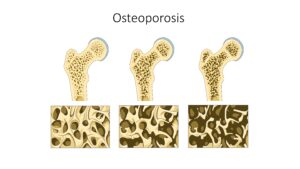
`Image by Google Image credit-https://images.app.goo.gl/xzY4yvs2pZg3Rzau5 - Osteoarthritis: A degenerative joint disease that can affect the joints of the appendicular skeleton especially the distal interphalangeal joints, and hip joints. There are two types of Arthritis as follows:
Primary Osteoarthritis
Secondary Osteoarthritis - Rheumatoid Arthritis: Rheumatoid arthritis is an autoimmune disorder that can affect multiple joints, including those in the appendicular skeleton. It is an inflammatory disorder characterized by Symmetrical peripheral polyarthritis. It leads to inflammation, joint pain, and damage to the affected joints.
- Stress Fractures: Stress fractures are hairline cracks in bones that result from repetitive stress over the bone surface. They are common in the lower extremities, especially in runners and athletes.
- Congenital Abnormalities: Some individuals are born with congenital abnormalities in the appendicular skeleton, such as hip dysplasia, or congenital limb deformities.
Treatment and management of these disorders of the appendicular skeleton can vary depending on the specific condition and level of severity. It may involve conservative measures like physical therapy, medication, or in some cases, surgical intervention. If suspected or diagnosed with a disorder of the appendicular skeleton, it’s essential to consult with a healthcare professional for a proper evaluation and treatment plan.